Arteries In Neck Diagram : This diagram depicts neck arteries and explains the details of neck arteries.. They can be called the main arteries of the head and neck. Instant anatomy is a specialised web site for you to learn all about human anatomy of the body with diagrams podcasts and revision q. The carotids reside beneath the skin on either side, and the pulse can be felt easily with your hand. Carotis communis doubles left longer than the right located on the neck behind the sternocleidomastoid. This diagram of the human heart shows all the major vessels, and arrows indicate the direction of flow through the heart.
Hold the position for as long as you can. The easiest spot is where it joins your head, just under the corner of the mandible. Blank neck diagrams help you memorize the fretboard. This diagram depicts neck arteries and explains the details of neck arteries. Rarely artery ascends in the neck without undergoing division, either eca or ica being absent.
Vascular Cat: Cat Veins - Cartoon Diagram from 4.bp.blogspot.com Quotes and sayings about death. They can be called the main arteries of the head and neck. Veins and arteries of the neck. This diagram of the human heart shows all the major vessels, and arrows indicate the direction of flow through the heart. Start studying arteries of neck. The distal part of the subclavian artery can be located as it emerges between the anterior and middle scalene muscles. The carotid artery pulse can be felt by pushing lateral to the upper border of the thyroid cartilage just under the anterior edge of the sternomastoid muscle. The left and right carotids, and the left and right vertebral arteries.
A neck swelling can also occur as accumulation of fluid, lymph, or inflammatory, or tumor cells in an area unde… this diagrams shows the major arteries in the human body.
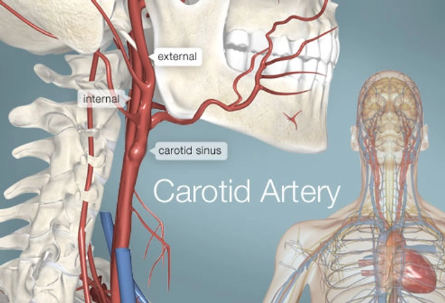
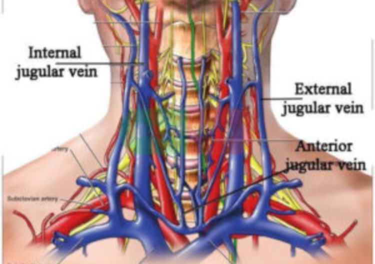
The neck diagram above shows you the structure and anatomy of the neck. Arteries in the neck picture. Instant anatomy is a specialised web site for you to learn all about human anatomy of the body with diagrams podcasts and revision q. There are 4 main arteries in your neck; Printable neck diagrams to help you learn more about the system that makes up our neck. Hold the position for as long as you can. Teeth diagram skeleton art print vintage anatomy art print on tea stained paper dog art dog s xmas for momdog christmas gift. Blank neck diagrams help you memorize the fretboard. The neck arteries 3 pages 1918 human anatomy by mysunshinevintage. They can be called the main arteries of the head and neck. The head and neck receives the majority of its blood supply through the carotid and vertebral arteries. In this image, you will find internal carotid artery, inferior thyroid vein, anterior jugular vein, suprascapular artery, dorsal scapular artery, superficial cervical artery, inferior jugular vein in it. Diagram of human heart :
You can use highlighters in different colors to see which notes are in a major scale, a minor scale, or a. Start studying arteries of neck. Brain diagram brain anatomy anatomy and physiology human anatomy carotid artery human anatomy picture definition conditions more. It runs from the heart down the length of the chest and abdomen. They can be called the main arteries of the head and neck.
Carotid Artery (Human Anatomy): Picture, Definition ... from img.webmd.com The neck diagram above shows you the structure and anatomy of the neck. Learn vocabulary, terms and more with flashcards, games and other study tools. The descending aorta is the largest artery in the body; Carotis communis doubles left longer than the right located on the neck behind the sternocleidomastoid. Bodytomy provides a labeled iliac artery diagram to help you understand the anatomy and function of the common iliac. Schematic owchart from the arteries in the neck and head. Hold the position for as long as you can. Rarely artery ascends in the neck without undergoing division, either eca or ica being absent.
Blank neck diagrams help you memorize the fretboard.
The internal carotid artery (latin: In this image, you will find internal carotid artery, inferior thyroid vein, anterior jugular vein, suprascapular artery, dorsal scapular artery, superficial cervical artery, inferior jugular vein in it. The external carotid artery supplies the areas of the head and neck external to the start studying arteries of head and neck. The distal part of the subclavian artery can be located as it emerges between the anterior and middle scalene muscles. There are 2 common carotid arteries: The easiest spot is where it joins your head, just under the corner of the mandible. 20 5 circulatory pathways anatomy and physiology. It carries blood from the left ventricle to the coronary arteries. This diagram depicts neck arteries and explains the details of neck arteries. Blank neck diagrams help you memorize the fretboard. Quotes and sayings about death. Arteries in the neck picture. Arteria carotis interna) is located in the inner side of the neck in contrast to the external carotid artery.
In the following diagrams, the anatomy is drawn with and without the labels. In human anatomy, they arise from the common carotid arteries where these bifurcate into the internal and external carotid arteries at cervical vertebral level 3 or 4. Quotes and sayings about death. The neck diagram above shows you the structure and anatomy of the neck. It commences in the substance of the parotid gland, on a level with the angle of the mandible, and runs perpendicularly down the neck, in the direction of a line drawn from the angle of the mandible to the middle of the clavicle at the posterior border of the sternocleidomastoideus.
Medical Axioms on Twitter: "By what magical mechanism ... from pbs.twimg.com Diagram of human heart : In the following diagrams, the anatomy is drawn with and without the labels. Posted by cassidy smith on 9 may 2018, 11:14 am. Ploaded with beautifully illustrated diagrams clearly and concisely labeled for easy identification. Blank neck diagrams help you memorize the fretboard. The internal carotid artery (latin: The cervical plexus supplies the skin and muscles of the anterolateral neck, the superior thorax, and an area of the scalp. In this image, you will find internal carotid artery, inferior thyroid vein, anterior jugular vein, suprascapular artery, dorsal scapular artery, superficial cervical artery, inferior jugular vein in it.
Neck diagram of muscles, arteries, and skeleton.
Printable neck diagrams to help you learn more about the system that makes up our neck. Internal jugular, external jugular cranial nerves (diagram). The distal part of the subclavian artery can be located as it emerges between the anterior and middle scalene muscles. Carotis communis doubles left longer than the right located on the neck behind the sternocleidomastoid. Neck diagram of muscles, arteries, and skeleton. The external carotid artery reduces in size while moving up the neck, giving various branches along the way. The carotids reside beneath the skin on either side, and the pulse can be felt easily with your hand. Blank neck diagrams are important to musicians for several reasons. Arteria carotis interna) is located in the inner side of the neck in contrast to the external carotid artery. In human anatomy, they arise from the common carotid arteries where these bifurcate into the internal and external carotid arteries at cervical vertebral level 3 or 4. Learn vocabulary, terms and more with flashcards, games and other study tools. The neck diagram above shows you the structure and anatomy of the neck. You can use highlighters in different colors to see which notes are in a major scale, a minor scale, or a.
Hold the position for as long as you can arteries in neck. The carotid artery pulse can be felt by pushing lateral to the upper border of the thyroid cartilage just under the anterior edge of the sternomastoid muscle.


0 Comments